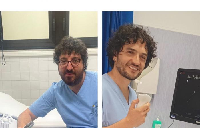
POTENZA – “The international journal ‘Pulmonology’ recently published the results of the ‘Ecovita’ study, the result of the collaboration of the San Carlo hospital in Potenza, the San Giovanni di Dio hospital in Melfi and 8 other Italian centers”. This was announced by the general director of the Aor San Carlo Giuseppe Spera, highlighting “the important scientific contribution of Dr. Luciano Restivo, of Acceptance and Emergency Medicine and Surgery of the Melfi hospital, and of Dr. Carlo Acierno, of Diseases Infectious diseases of San Carlo di Potenza, who used emergency chest ultrasound in patients with severe acute respiratory syndrome associated with Coronavirus-2, during the dark pages of the pandemic emergency”.
“Application and internal validation of lung ultrasound score in COVID-19 setting: the ECOVITA observational study” is the title of the publication, already indexed in the international bibliographic archive PubMed. This is a prospective multicenter observational study, led by Professor Luca Rinaldi of the University of Molise, applied on 1,007 adult patients treated in 10 Italian centers between February and July 2021.
The objective of the study is to validate chest zonation in 12 fields of application of the ultrasound probe, assigning a variable numerical index based on the presence or absence of pulmonary densifications.
“The validation of the 12-zone LUS (thoracic ultrasound) score – explains Dr. Restivo – is aimed at offering a parameter for predicting the severity of the COVID 19 disease in terms of mortality and the use of invasive ventilatory support. To our knowledge, the Ecovita study is the first that aimed to validate the LUS score in such a large patient population.”
“The severity of the pathology is mainly linked to the involvement of the lower airways – continue doctors Acierno and Restivo – resulting in pneumonia, respiratory failure, acute respiratory distress syndrome (ARDS). Chest CT has represented the gold standard in the diagnostic definition and evaluation of the extent of lung pathology linked to Coronavirus 19, however, considering the exposure to ionizing radiation, the commitment of time and resources, the difficulty in transporting patients from the treatment departments to the Radiology Units, it was necessary to validate and strengthen an alternative method, easy to use, reproducible, non-toxic, non-invasive, such as thoracic ultrasound”.
The ultrasound study of the chest is a relatively recent subject compared to the decades-long history of ultrasound, however, the extensive use of LUS during the pandemic has led to an increase in acquisitions and evidence for which it is now universally accepted as a diagnostic method.
The Ecovita study confirms that thoracic ultrasound is a reproducible, inexpensive and non-toxic technique, useful for assessing the severity of Sars-CoV-2 disease and offering precise risk stratification. LUS, in fact, accurately predicted mortality and severity of the clinical course of COVID patients.


